Datei:Unctional complex and pinocytotic vesicles - embryonic brain - TEM.jpg

Originaldatei (1.600 × 1.278 Pixel, Dateigröße: 861 KB, MIME-Typ: image/jpeg)
Beschreibung
| Beschreibung |
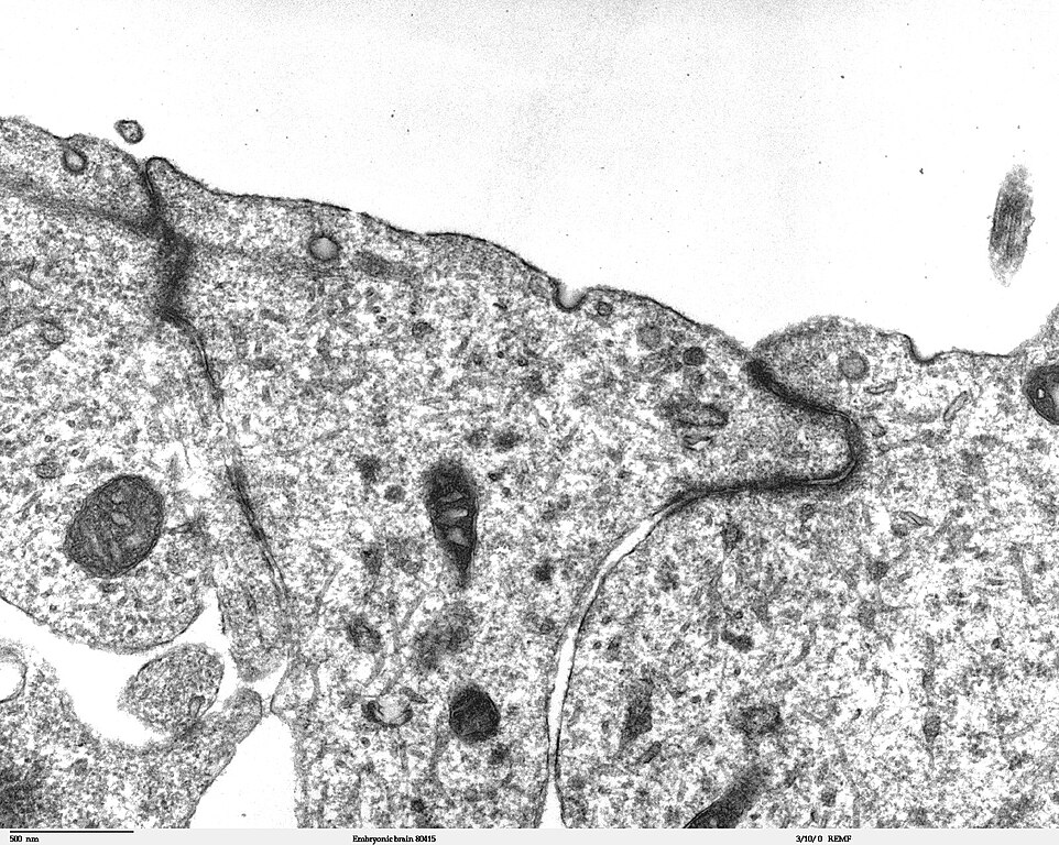
Transmission electron microscope image of a thin section cut through the developing brain tissue (telencephalic hemisphere) of an 11.5 day mouse embryo. This image of the luminal surface of the telencephalon, shows junctional complexes and pinocytotic vesicles. The junctional complex is divided into three types of junctions: 1) the most apical is the tight junction, which controls and/or restricts the movement of molecules across epithelial layers and helps maintain polarity, 2) the zonula adherens, which also includes the numerous actin filaments seen in the apical cytoplasm, and 3) the desmosome, which is a spot junction. The pinocytotic vesicles are formed from coated pits in the plasma membrane and are involved in endocytosis. JEOL 100CX TEM References: Marin-Padilla, M. (1985) "Early Vascularization of the Embryonic Cerebral Cortex: Golgi and Electron Microscope Studies", J. Comparative Neurology, 241:237-249 Marin-Padilla, M. and M. Amievo (1989) "Early Neurogenesis of the Mouse Olfactory Nerve: Golgi and Electron Microscope Studies", J. Comparative Neurology, 288:339-352 |
| Quelle | |
| Urheber | Louisa Howard, Miguel Marin-Padilla |
| Genehmigung (Weiternutzung dieser Datei) |
PD |
Lizenz
| Dieses Werk wurde von seinem Urheber Louisa Howard, Miguel Marin-Padilla als gemeinfrei veröffentlicht. Dies gilt weltweit. In manchen Staaten könnte dies rechtlich nicht möglich sein. Sofern dies der Fall ist: Louisa Howard, Miguel Marin-Padilla gewährt jedem das bedingungslose Recht, dieses Werk für jedweden Zweck zu nutzen, es sei denn, Bedingungen sind gesetzlich erforderlich.
|
Dateiversionen
Klicke auf einen Zeitpunkt, um diese Version zu laden.
| Version vom | Vorschaubild | Maße | Benutzer | Kommentar | |
|---|---|---|---|---|---|
| aktuell | 21:06, 2. Nov. 2006 | | 1.600 × 1.278 (861 KB) | wikimediacommons>Patho | {{Information |Description=Transmission electron microscope image of a thin section cut through the developing brain tissue (telencephalic hemisphere) of an 11.5 day mouse embryo. This higher magnification image of "Embryonic brain 80415", shows an area o |
Dateiverwendung
Keine Seiten verwenden diese Datei.